Tumors of the bile duct and gallbladder
Bile duct and gallbladder tumors, both noncancerous and cancerous, are rare.
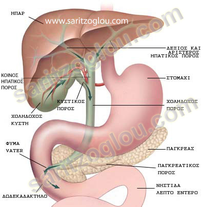
Bile is a fluid produced by the liver that helps with digestion. It is carried through small tubes (bile ducts) that carry bile through the liver and then from the liver to the gallbladder and small intestine. The gallbladder is a small, pear-shaped sac located under the liver that stores bile and releases it when needed.
Bile duct cancer (cholangiocarcinoma) is rare. It can arise anywhere in the bile ducts, especially in the bile ducts that lie outside the liver.
Age and primary sclerosing cholangitis increase the risk of developing this cancer.
Gallbladder cancer is also rare. Almost everyone with gallbladder cancer has gallstones. Many people live only a few months after developing this cancer.
This cancer is more common among American Indians, people with large gallstones, and people with extensive scarring of the gallbladder, which can occur in severe chronic cholecystitis.
Polyps, which are noncancerous (benign) growths of tissue, can grow in the gallbladder. They rarely cause symptoms or require treatment. They are found in about 5% of people during an ultrasound. Surgery may be needed to remove larger polyps.
Sometimes cancers can block the flow of bile, but most blockages are caused by gallstones. Even less often, cancer can spread (metastasis) from elsewhere in the body to nearby structures or nearby lymph nodes, causing a blockage. Noncancerous tumors in the bile ducts also cause blockages.
Bile Duct and Gallbladder Tumors – Symptoms
- Worsening jaundice (yellowish discoloration of the skin and whites of the eyes)
- Abdominal discomfort
- Loss of appetite
- Weight loss
- Itching
- Feeling tired.
The symptoms gradually get worse.
Abdominal pain may become increasingly severe and constant. The pain is usually caused by a blockage of the bile ducts. Stools may be light-colored.
Most gallbladder polyps do not cause symptoms.
Diagnosis
Bile duct or gallbladder cancer is suspected when a bile duct is blocked and no other cause is found.
Bile duct cancer is suspected especially in people with primary sclerosing cholangitis (PSC).
The diagnosis is confirmed by imaging tests.
Usually, an ultrasound is done first.
Sometimes a computed tomography (CT) scan is done, but the results are often inconclusive.
CT cholangiography (a computed tomography scan of the bile ducts done after an injection of a contrast agent into a vein) or magnetic resonance cholangiopancreatography (MRCP) is usually needed.
If the results of imaging tests are unclear, an endoscopic retrograde cholangiopancreatography (ERCP) is done.
In this procedure, a flexible, lighted tube (endoscope) is inserted through the mouth and into the small intestine. A thin tube (catheter) is inserted through the endoscope, and a radiopaque contrast agent, which is visible on X-rays, is injected through the catheter into the bile ducts. X-rays are then taken to look for any abnormalities.
This procedure allows doctors to obtain images and a tissue sample for examination under a microscope.
If these tests suggest a tumor but are not definitive, doctors may take a tissue sample by inserting a thin needle through the skin into the area that is thought to be abnormal. Ultrasound or CT scan is used to guide the needle.
To determine how far the cancer has spread, surgery may be needed to directly examine the area (a procedure called diagnostic laparoscopy or open laparotomy).
Bile Duct and Gallbladder Tumors – Treatment
Stents in Blocked Bile Ducts
Sometimes surgery to remove the tumor
Most bile duct and gallbladder cancers are fatal, but treatment can help control symptoms.
Tubes (stents) inserted into a duct allow bile to flow past the blockage. This procedure helps control pain and relieves itching. Stents can be placed during endoscopic retrograde cholangiopancreatography (ERCP).