Intestinal Obstruction
An intestinal obstruction is a condition that completely stops or severely affects the passage of food, fluids, digestive secretions, and gas through the intestines.
The most common causes in adults are scar tissue resulting from previous abdominal surgery, hernias, and tumors.
Pain, bloating, and loss of appetite are common.
Diagnosis is based on the results of a physical exam and X-rays.
Surgery is often needed to remove the obstruction.
A blockage can occur anywhere along the small or large intestine and can be partial or complete. The part of the intestine above the blockage continues to function. This part of the intestine enlarges as it fills with food, fluids, digestive secretions, and gas. The intestinal lining swells and becomes inflamed. If the condition is left untreated, the intestine can rupture, leaking its contents and causing inflammation and infection of the abdominal cavity (peritonitis).
Intestinal Obstruction – Causes
The causes of intestinal obstruction vary depending on the age of the person and the location of the obstruction.
In newborns and infants, intestinal obstruction is usually caused by a genetic defect, a hard mass of intestinal contents (meconium plug syndrome), a twisting of a loop of intestine (volvulus), a narrowing or absence of a section of intestine (intestinal atresia), or telescoping of one section of intestine into another (intussusception).
In adults, the most common causes overall are bands of internal scar tissue resulting from previous abdominal surgery (adhesions), parts of the intestine that bulge through an abnormal opening (hernias), and tumors. The likelihood of a specific cause varies depending on the part of the intestine affected.
A blockage of the first part of the small intestine (duodenum) can be caused by pancreatic cancer, scarring from an ulcer, or Crohn’s disease. Rarely, a gallstone or a mass of undigested food can block other parts of the small intestine.
A blockage of the large intestine is usually caused by cancer, diverticulitis, or a hard mass of stool (fecal impaction). Adhesions and abscesses are less common causes of large bowel obstruction.
Strangulation
If an obstruction cuts off the blood supply to the intestine, the condition is called strangulation. Strangulation occurs in about 25% of people with small bowel obstruction. Strangulation usually occurs when part of the intestine becomes trapped in an abnormal opening (strangulated hernia), abscess, or intussusception. Gangrene can develop in as little as 6 hours. With gangrene, the intestinal wall dies, usually causing a rupture, which leads to peritonitis, septic shock, and, if left untreated, death.
What causes intestinal strangulation?
Intestinal strangulation (cut-off of blood supply to the intestine) usually results from one of three causes: strangulated hernia, volvulus, or intussusception.
Symptoms
Symptoms of intestinal obstruction usually include cramping pain in the abdomen, bloating, and loss of appetite. The pain tends to come in waves and eventually becomes continuous. Vomiting is common with small bowel obstruction, but is less common and begins later with large bowel obstruction.
Complete obstruction causes severe constipation, while partial obstruction can cause diarrhea.
With strangulation, the pain can be severe and constant. Fever is common and is especially likely if the intestinal wall is torn.
With volvulus (twisting), the pain often begins abruptly.
Intestinal Obstruction – Diagnosis
- Examination of the abdomen by a doctor
- X-rays
- Computed tomography (CT) scan
A doctor examines the abdomen for tenderness, swelling, or masses. When an obstruction occurs, the abdomen is almost always swollen. The sounds normally made by a functioning bowel (bowel sounds), which can be heard through a stethoscope, may be much louder, higher-pitched, or absent. The abdomen is usually not very tender when the doctor presses on it unless the rupture has caused peritonitis.
Imaging of the abdomen, such as X-rays or CT scans, is usually done.
X-rays may show enlarged loops of bowel that indicate the location of the obstruction. They may also show air around the intestine or under the layer of muscle that separates the abdomen and chest (diaphragm). Air is not usually found in these places and is therefore a sign of a ruptured intestine.
An abdominal CT scan is often used to get a better picture of the intestines and to pinpoint the exact location and cause of the blockage.
Intestinal Obstruction – Treatment
- Suction through a nasogastric tube
- Fluids given through a vein
- Surgery for strangulation
- Sometimes a colostomy
- Someone suspected of having an intestinal obstruction is hospitalized.
Typically, a long, thin tube is passed through the nose and placed into the stomach (called a nasogastric tube) or intestine. Suction is applied to the tube to remove material that has accumulated above the blockage. Fluids and electrolytes are given through a vein to replace water and salts lost from vomiting or diarrhea.
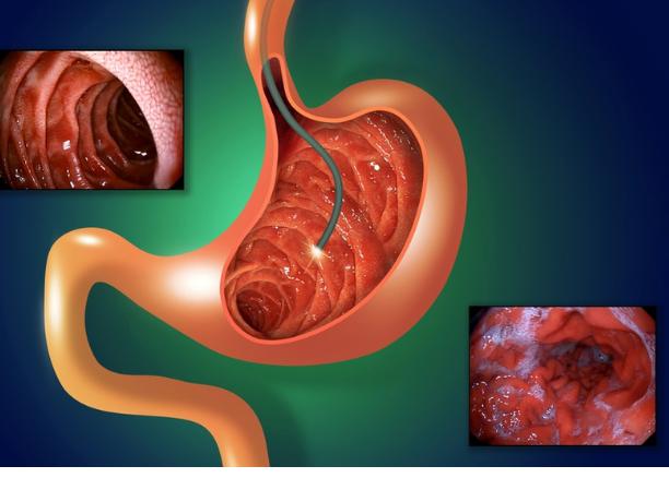
Sometimes an obstruction resolves without further treatment, especially if it is caused by scarring or adhesions. Occasionally, an endoscope (a flexible viewing tube) inserted through the anus, or a barium enema, which distends the colon, may be used to treat certain disorders, such as a twisted segment of bowel in the lower part of the colon.
Most often, however, surgery is done as soon as possible if doctors are concerned about strangulation.
The cause of the obstruction and the appearance of the bowel determine whether the surgeon can relieve the obstruction without removing a section of bowel.
Sometimes adhesions can be cut to release the trapped section of bowel, but they can recur, and the obstruction can recur.
In some cases, an ileostomy (a procedure in which the cut end of the small intestine is permanently connected to a surgical opening in the abdominal wall) or colostomy (a surgically created opening between the large intestine and the abdominal wall) is needed to relieve a blockage.
Understanding Colostomy
In a colostomy, the large intestine (colon) is cut. The healthy end of the large intestine, which was before the blockage, is brought to the surface of the skin through an opening surgically created in the abdominal wall. It is then sewn to the skin of the opening. Stool passes through the opening and into a disposable bag. The colostomy allows the remaining part of the large intestine to rest while the person recovers. After the person recovers from surgery and the colon heals, the two ends can be reconnected so that stool can pass normally.